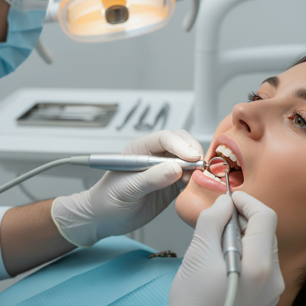
Teeth filling represents a fundamental restorative dental procedure that addresses cavitated lesions, structural damage, and tooth decay through material replacement and rehabilitation. This evidence-based treatment modality preserves compromised tooth structure while restoring biomechanical function and aesthetic continuity.
Clinical Indications for Dental Filling Procedures
Pathophysiology of Dental Caries
Dental caries develop through a complex demineralization process initiated by acidogenic bacteria, primarily Streptococcus mutans and Lactobacillus species. These microorganisms metabolize fermentable carbohydrates, producing organic acids that dissolve hydroxyapatite crystals in tooth enamel.
When demineralization exceeds remineralization capacity, cavitation occurs, requiring mechanical intervention to remove infected dentin and restore tooth integrity. The progression from initial enamel lesions to deep dentinal caries determines treatment complexity and material selection.
Structural Tooth Damage Requiring Restoration
Beyond bacterial decay, teeth fillings address various structural deficiencies:
- Traumatic fractures compromising tooth morphology
- Abrasion and attrition from mechanical wear
- Erosion from dietary acids or gastric reflux
- Developmental defects including enamel hypoplasia
- Failed or deteriorating previous restorations
Classification of Restorative Filling Materials
Resin-Based Composite Systems
Composite resin materials consist of an organic polymer matrix (typically bis-GMA or UDMA) reinforced with inorganic filler particles. Contemporary formulations achieve 70-80% filler loading by weight, providing enhanced mechanical properties and wear resistance.
Material characteristics:
- Micromechanical and chemical bonding to tooth structure via adhesive systems
- Polymerization shrinkage of 1.5-3% volume reduction
- Coefficient of thermal expansion similar to natural tooth structure
- Compressive strength of 250-300 MPa
- Flexural strength of 80-120 MPa
- Clinical longevity of 5-7 years in posterior applications
Clinical advantages:
- Superior aesthetic integration through polychromatic layering techniques
- Minimal invasive preparation requirements due to adhesive bonding
- Immediate load-bearing capacity post-polymerization
- Versatility for direct and indirect fabrication methods
Material limitations:
- Technique-sensitive placement requiring moisture control
- Polymerization shrinkage stress potentially causing marginal gap formation
- Reduced wear resistance compared to amalgam in high-stress zones
- Higher material costs and chair time requirements
Dental Amalgam Alloys
Amalgam represents a time-tested restoration material combining approximately 50% elemental mercury with silver-tin-copper alloy powders. High-copper amalgam formulations (>12% copper) demonstrate superior corrosion resistance and mechanical properties.
Physical and mechanical properties:
- Compressive strength exceeding 380 MPa
- Excellent wear resistance in occlusal stress-bearing areas
- Clinical survival rates of 10-15 years or greater
- Self-sealing corrosion products at restoration margins
- Minimal technique sensitivity during placement
Safety profile:
- Mercury exposure from dental amalgam remains below established safety thresholds
- No demonstrable systemic health effects in general populations
- Dental regulatory bodies worldwide affirm amalgam safety for most patients
- Environmental concerns regarding mercury disposal require proper waste management
Contemporary usage considerations:
- Declining utilization due to aesthetic concerns and patient preferences
- Potential galvanic reactions when contacting other metallic restorations
- Thermal conductivity necessitating adequate dentinal insulation
- Macromechanical retention requires additional tooth structure removal
Ceramic and Glass-Ceramic Restorations
Ceramic materials encompass feldspathic porcelain, leucite-reinforced ceramics, and lithium disilicate glass-ceramics for indirect restorations. These biocompatible materials exhibit exceptional aesthetic properties and chemical stability.
Material advantages:
- Highest aesthetic potential with natural translucency and opalescence
- Superior color stability and stain resistance
- Excellent biocompatibility with minimal allergic potential
- Flexural strength ranging from 120-400 MPa depending on composition
- Clinical longevity approaching 15 years for bonded restorations
Application specifications:
- Primarily utilized for inlay, onlay, and veneer fabrications
- Requires adhesive cementation protocols for optimal performance
- Laboratory or CAD/CAM fabrication methods
- Higher cost structure compared to direct restoration techniques
Glass Ionomer Cements
Glass ionomer materials chemically bond to tooth structure through ionic exchange with hydroxyapatite. These self-adhesive materials release fluoride ions, providing cariostatic effects in high-risk patients.
Clinical applications:
- Pediatric dentistry for primary teeth restorations
- Atraumatic restorative technique (ART) applications
- Cervical and root surface lesions
- Core build-up materials and temporary restorations
- Liner or base materials under composite restorations
Material considerations:
- Lower mechanical strength limits use in stress-bearing areas
- Moisture sensitivity during setting reaction
- Continuous fluoride release for secondary caries prevention
- Resin-modified formulations offer improved physical properties
Noble Metal Alloys
Gold and gold alloy restorations demonstrate unparalleled longevity and biocompatibility. Cast gold restorations can function for 20+ years with appropriate oral hygiene maintenance.
Clinical characteristics:
- Exceptional marginal adaptation and seal
- Minimal wear of opposing dentition
- Excellent corrosion resistance
- Precise castability for complex preparations
- Prohibitive cost and aesthetic limitations restrict current usage
Evidence-Based Filling Procedure Protocol
Pre-operative Assessment and Diagnosis
Comprehensive examination incorporates:
- Visual and tactile inspection using sharp explorers
- Radiographic evaluation (bitewing and periapical imaging)
- Transillumination and optical caries detection devices
- Assessment of pulpal vitality through thermal and electric testing
- Caries risk evaluation and preventive protocol implementation
Anesthesia Administration
Local anesthetic infiltration or nerve block techniques provide pulpal and soft tissue anesthesia. Common agents include:
- Articaine hydrochloride 4% with epinephrine 1:100,000
- Lidocaine hydrochloride 2% with epinephrine 1:100,000
- Mepivacaine hydrochloride 3% (vasoconstrictor-free formulation)
Anesthetic onset occurs within 3-5 minutes with duration of 45-90 minutes for infiltration techniques. Complete pulpal anesthesia confirmation precedes operative procedures.
Caries Excavation and Cavity Preparation
Excavation principles:
- Complete removal of infected, non-remineralizable dentin
- Preservation of affected but remineralizable dentin
- Maintenance of pulpal health through conservative preparation depth
- Establishment of mechanical retention features (amalgam) or enamel margin beveling (composite)
Modern minimally invasive dentistry emphasizes maximum preservation of sound tooth structure. Chemomechanical caries removal systems and polymer burs enable selective excavation techniques.
Adhesive Bonding Protocols for Composite Restorations
Etch-and-rinse technique:
- 35-37% phosphoric acid etchant application for 15 seconds on enamel, 10 seconds on dentin
- Thorough water rinse and gentle air drying to moist dentin
- Primer and adhesive resin application with light polymerization
Self-etch adhesive systems:
- Acidic primer simultaneously conditions and primes tooth structure
- Simplified application reducing technique sensitivity
- Decreased post-operative sensitivity in some clinical scenarios
Incremental Composite Placement
Composite resin is placed in 2mm increments to manage polymerization shrinkage stress. Each increment undergoes light polymerization for 20-40 seconds depending on material opacity and curing light intensity.
Placement strategy:
- Initial dentin replacement increment
- Intermediate body layers establishing bulk contours
- Final enamel-shade increment for aesthetic surface characterization
- Occlusal morphology recreation including cuspal inclines and developmental grooves
Occlusal Adjustment and Finishing
Articulating paper identifies premature contacts requiring selective adjustment. High-speed finishing diamonds and carbide burs refine contours followed by aluminum oxide discs and polishing systems for surface smoothness.
Proper finishing reduces plaque retention and enhances restoration longevity. Final occlusal verification ensures balanced contacts in centric and excursive movements.
Clinical Benefits of Early Restorative Intervention
Maximum Tooth Structure Conservation
Prompt cavity restoration when lesions remain confined to enamel or superficial dentin requires minimal tooth reduction. Since enamel cannot regenerate, preservation of maximum tooth structure maintains biomechanical integrity and long-term prognosis.
Delayed treatment necessitates increasingly extensive preparations as caries progresses apically and laterally. Large restorations compromise tooth strength and may require cuspal coverage with indirect restorations.
Simplified Treatment Modalities
Early cavity detection allows straightforward direct restoration placement. Advanced lesions approaching or involving the pulp require endodontic therapy followed by post-and-core reconstruction and full-coverage crowns.
The procedural complexity escalation directly correlates with treatment costs and appointment requirements. Minimally invasive early intervention represents optimal clinical practice.
Prevention of Pulpal Pathology
Bacterial penetration into deep dentin tubules and pulpal exposure results in irreversible pulpitis requiring root canal treatment. Early restoration eliminates bacterial reservoirs before pulpal involvement occurs.
Untreated apical periodontitis can develop into acute dental abscesses with potential for serious systemic complications including Ludwig’s angina, cavernous sinus thrombosis, and bacterial endocarditis in susceptible patients.
Economic Optimization
Cost-effectiveness analysis demonstrates significant savings through preventive and early restorative care. Australian fee survey data indicates:
- Simple composite restoration: $200-$300
- Root canal therapy with crown: $2,500-$4,500
- Implant-supported crown: $4,000-$7,000
Early intervention provides 85-90% cost reduction compared to complex rehabilitative procedures.
Post-Operative Sequelae and Management
Transient Dentinal Hypersensitivity
Post-restorative sensitivity represents the most common complication, affecting 5-30% of patients depending on cavity depth and restoration type.
Temporal progression:
- Peak sensitivity: 24-48 hours post-operatively
- Gradual resolution: 7-14 days for most cases
- Extended sensitivity: 2-4 weeks for deep preparations or large restorations
Etiological factors:
- Residual inflammatory response in odontoblastic processes
- Microleakage at restoration-tooth interface
- Inadequate polymerization or pulpal insulation
- High occlusal contacts causing mechanical irritation
- Postoperative pulpal hyperemia
Clinical Management Protocols
Pharmacological interventions:
- NSAIDs (ibuprofen 400-600mg) for inflammatory component
- Desensitizing toothpaste containing potassium nitrate or stannous fluoride
- Topical fluoride varnish application at restoration margins
Behavior modification:
- Avoidance of thermal extremes during initial healing period
- Mastication on contralateral side for 24-48 hours
- Gentle oral hygiene maintenance without excessive force
Indications for Clinical Re-evaluation
Persistent or worsening symptoms beyond expected timelines warrant professional assessment:
- Pain intensification after 48-hour mark
- Sharp, reproducible pain on biting suggesting occlusal interference
- Prolonged sensitivity exceeding 4-week duration
- Spontaneous pain without external stimuli
- Visible restoration fracture or marginal breakdown
These symptoms may indicate reversible pulpitis requiring occlusal adjustment, restoration replacement, or irreversible pulpitis necessitating endodontic intervention.
Evidence-Based Maintenance Strategies
Professional Monitoring Protocols
Systematic restoration evaluation during recall examinations identifies potential failures before symptom development:
- Visual inspection for marginal discoloration or ditching
- Explorer tactile examination for surface defects
- Radiographic assessment for secondary caries or marginal gaps
- Occlusal evaluation for wear patterns and contacts
- Periodontal probing around restoration margins
Patient-Centered Preventive Measures
Mechanical biofilm control:
- Twice-daily brushing with fluoridated dentifrice (1450 ppm fluoride minimum)
- Daily interdental cleaning using floss or interdental brushes
- Consideration of powered toothbrushes for enhanced plaque removal efficacy
Chemical adjuncts:
- Fluoride mouthrinse for high caries-risk patients
- Chlorhexidine gluconate for enhanced antimicrobial control
- Xylitol-containing products for bacterial metabolism interference
Dietary modification:
- Limitation of fermentable carbohydrate frequency and duration
- pH-neutral beverage selection to minimize erosive potential
- Adequate hydration maintaining salivary flow and buffering capacity
Cost Analysis for Australian Dental Market
Restoration costs vary significantly based on material selection, geographic location, and practice fee structures.
2024-2025 fee survey data:
| Restoration Type | Size Category | Average Cost Range (AUD) |
| Amalgam | Small (1 surface) | $150-$200 |
| Amalgam | Large (3+ surfaces) | $250-$400 |
| Composite Resin | Small (1 surface) | $200-$270 |
| Composite Resin | Large (3+ surfaces) | $300-$500 |
| Ceramic Inlay/Onlay | N/A | $145-$395 |
| Gold Restoration | Variable | $300-$1,000+ |
Private health insurance typically provides 50-80% coverage for basic restorative procedures depending on policy level and annual limits. Some practices offer payment plan arrangements for patients without insurance coverage.
Specialized Restorative Services
At Dental Smiles, evidence-based teeth filling services utilize contemporary materials and minimally invasive techniques. The practice emphasizes:
- Biocompatible tooth-colored composite systems for aesthetic zones
- Efficient single-visit direct restoration protocols
- Comprehensive caries risk assessment and preventive counseling
- Immediate access for acute dental emergencies
- Transparent fee structures and flexible payment options
Detailed service information is available through the teeth filling service page for patients in the Charmhaven and Chester Hill service areas.
Clinical Decision-Making Framework
Optimal restoration material selection requires integration of multiple variables:
Patient factors:
- Aesthetic expectations and smile zone considerations
- Caries risk classification and oral hygiene capacity
- Occlusal forces and parafunctional habits
- Allergy history and material biocompatibility
- Financial constraints and insurance coverage
Tooth-specific factors:
- Cavity size, location, and remaining tooth structure
- Pulpal proximity and vitality status
- Strategic importance and restorability assessment
- Adjacent and opposing tooth conditions
Evidence-based considerations:
- Material longevity data from longitudinal clinical studies
- Failure mode analysis and replacement requirements
- Cost-effectiveness ratios over restoration lifespan
- Contemporary adhesive technology capabilities
Shared decision-making incorporating patient preferences with clinical expertise optimizes treatment outcomes and satisfaction.
Contemporary Trends in Restorative Dentistry
Minimally invasive dentistry philosophy dominates modern practice patterns, emphasizing biological preservation over mechanical retention. Adhesive technologies enable conservative preparations maintaining maximum tooth structure.
Digital dentistry integration through intraoral scanning and CAD/CAM fabrication streamlines indirect restoration workflows. Same-visit ceramic restorations eliminate temporary restoration requirements and reduce appointment burden.
Bioactive restorative materials incorporating calcium and phosphate ion release demonstrate remineralization potential at restoration margins. These materials represent the convergence of restorative and preventive dentistry paradigms.
Artificial intelligence applications for caries detection and treatment planning show promise for enhanced diagnostic accuracy and standardized care protocols. Implementation of these technologies requires validation through rigorous clinical research.


